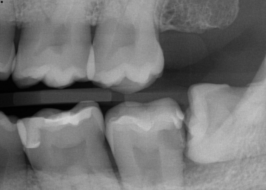
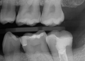
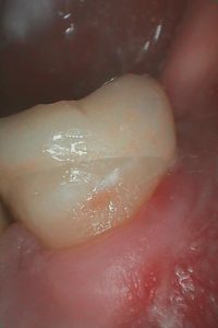
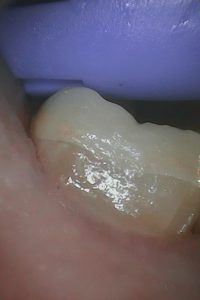
The horizontally positioned wisdom tooth (#17) caused decay on #18
Pt was referred to the oral surgeon to have #17 taken out
Pt was then seen by a root canal specialist (Dr. Bexter Yang) because the cavity was too deep
Greater Curve Technique was used to seal the deep cavity with tooth color filling
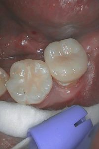
Final crown
Lower Molar with Deep/Long Cavity
Concerns: Second molar on the lower left side (#18) developed deep cavity on the root region due to the impacted wisdom tooth (#17).
Treatment: Extraction of #17 by an oral surgeon then root canal therapy of #18 by Dr. Bexter Yang. Porcelain crown with zirconia substructure on #18 to protect the remaining tooth structure.
A1. #17 (wisdom tooth) was impacted horizontally; decay is visible on #18 (second molar) on the x-ray (the shadow on the right side of the tooth). This is a very common decay because it is impossible for patient to reach and clean between #17/18.
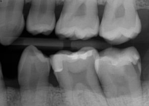
A2. X-ray of #18 after #17 was taken out. The bone level around #18 is not ideal, but since there is no mobility on #18 and the patient would really like to keep the tooth, patient was referred to the root canal specialist. Dr. Yang reviewed the case and deemed the tooth savable, so root canal therapy was performed.
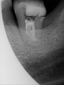
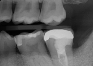
A3. X-ray of #18 after the root canal therapy (by Dr. Bexter Yang) with temporary filling.
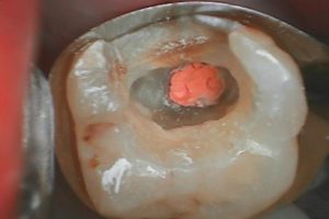
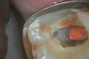
B1. #18 after the temporary filling and cavity is removed. The tooth was isolated by Greater Curve matrix band to create ideal seal.
B2. First layer of tooth color filling was placed to seal the edge of the tooth.
B3. X-ray was taken to check the seal.
C1. BruxZir (porcelain crown with zirconia substrate) was fabricated as final restoration. The x-ray was taken before cementation to check margin of the crown. The crown was done above the gum line because it is harder for the lab to make a crown margin as smooth as the one we did with tooth color filling. Rough margin will cause irritation of the gum, risk of calculus buildup, risk of caries.
C2.-C4. Different view of the final crown.

